Brain anatomy can be [[daunting]]. Let's try to tackle it step by step and start with the surfaces of the cerebral hemispheres. The surface of the cerebral hemispheres consists of many convolutions, the cortical gyri, which are separated by sulci of varying sizes. Even if you aced neuroanatomy during medical school and remember all cortical gyri and sulci by heart (which you probably don't:)), trying to find and recognize them on an MRI or CT-exam of the brain can seem like an impossible task at first. No worries, it isn't. With a little bit of practice and some tips and tricks, you'll be able to recognize the most important surface landmarks of the brain in no time. Let's start with the simplest and most important one: the central sulcus.
脑部解剖学可能令人望而生畏。让我们一步步来应对它,从大脑半球的外表面开始。大脑半球的外表面由许多褶皱组成,即皮质脑回,它们被不同大小的沟分隔。即使你在医学院期间神经解剖学学得很好,并且能记住所有的皮质脑回和沟(你可能做不到:)),在脑部 MRI 或 CT 检查中找到和识别它们可能一开始看起来是一项不可能的任务。别担心,这并不是。只要稍加练习和一些技巧,你很快就能识别出大脑最重要的表面标志。让我们从最简单和最重要的一个开始:中央沟。
The central sulcus is an important anatomic landmark on imaging. The central sulcus separates the frontal from the parietal lobe and is the sulcus between the precentral and the postcentralis gyrus, which constitute the primary motor cortex and primary somatosensory cortex respectively. The primary motor cortex is responsible for the control and execution of voluntary movements and lesions in the precentral gyrus will lead to a contralateral hemiparesis. The primary somatosensory cortex is responsible for the primary processing of somatic sensations.
中央沟是影像学上的一个重要解剖标志。中央沟将额叶与顶叶分开,是中央前回和中央后回之间的沟,分别构成初级运动皮层和初级躯体感觉皮层。初级运动皮层负责控制和执行随意运动,中央前回的损伤将导致对侧偏瘫。初级躯体感觉皮层负责躯体感觉的初级处理。
It's easiest to find the central sulcus in the axial plane. I've chosen an axial T2-weighted image for this tutorial, but you can find the central sulcus on any sequence off course. The central sulcus is easiest to detect relatively high up in the brain, above the level of the lateral ventricles.
在轴位平面上寻找中央沟最为容易。我在这个教程中选择了轴位 T2 加权图像,但当然你也可以在任何序列中找到中央沟。中央沟最容易检测到的位置相对较高,位于侧脑室上方。

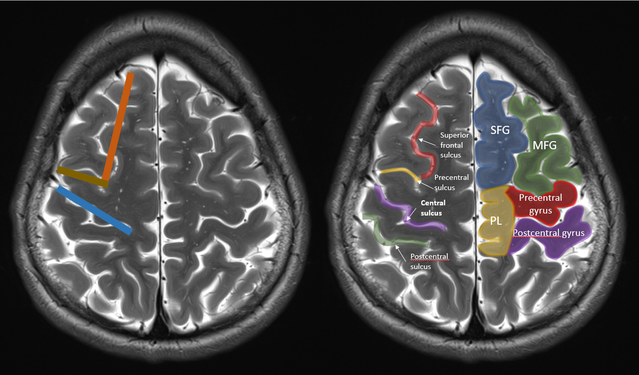
Let's start with the easiest one (in my opinion, almost never lets me down). Do you see the deep sulcus anteriorly in the brain? That's the superior frontal sulcus. I've coloured it red on the image below. The superior frontal sulcus separates the superior frontal gyrus (blue) from the middle frontal gyrus (green). At the posterior border of the middle frontal gyrus, the superior frontal sulcus (red) forms a juntion with the precentral sulcus (yellow). The combination of the superior frontal sulcus and the precentral sulcus looks a bit like capital letter “L” (or an inverted capital letter “L” depending on which hemisphere you're looking at).
让我们从最简单的一个开始(在我看来,几乎从未让我失望)。你能在脑部看到深沟吗?那就是额上沟。我在下面的图像中将其标记为红色。额上沟将额上回(蓝色)与中间额回(绿色)分开。在额中回的后缘,额上沟(红色)与中央沟(黄色)形成一个连接。额上沟和中央沟的组合看起来有点像大写字母“L”(或者取决于你观察的是哪个半球,像倒置的大写字母“L”)。

We're almost there! The gyrus behind the precentral sulcus is the precentral gyrus (red). The sulcus behind the precentral gyrus is the central sulcus (purple), the gyrus behind that the postcentral gyrus (purple), and the sulcus behind that the postcentral sulcus (green). Easy, no? We've found the central sulcus and it took us no effort at all. Just look for capital letter "L" and the central sulcus is the sulcus immediately behind the foot of letter "L".
我们几乎就找到了!位于中央沟后面的回是中央前回(红色)。中央前回后面的沟是中央沟(紫色),再后面的回是中央后回(紫色),再后面的沟是中央后沟(绿色)。简单吧?我们已经找到了中央沟,而且毫不费力。只需寻找大写字母“L”,中央沟就是位于字母“L”的底部后面的沟。

Some small details: notice that the central sulcus doesn't run all the way up to the medial hemispheric surface (sometimes it does, but most of the time it doesn't). As a consequence, there's not sulcus separating the pre- and postcentral gyrus at the medial surface of the brain. We call this part of the pericentral region "the paracentral lobule" and I've drawn it in yellow on the image above. The primary motor and somatosensory cortex are somatotopically organized and the paracentral lobule contains the motor (anterior) and somatosensory (posterior) representations of the contralateral leg. In other words: a hemorrhage or an infarction in the paracentral lobule, especially when anteriorly in the paracentral lobule, will cause a paresis of the contralateral leg. Also notice on the images above that in the left hemisphere, there's some kind of "cortical bridge" between the middle frontal gyrus and the precentral gyrus, with no precentral sulcus separating the two gyri. This is just one of many possible anatomic variations we might encounter when studying brain surface anatomy, and that's why we need some other tricks to recognize the central sulcus.
一些小细节:注意中央沟并没有一直延伸到大脑半球的内侧表面(有时会,但大多数时候不会)。因此,在大脑内侧表面并没有沟将中央前回和中央后回分开。我们将这部分中央区称为“中央旁小叶”,并在上图中用黄色标出。初级运动皮层和体感皮层是按照躯体定位组织的,旁中央小叶包含对侧腿的运动(前部)和体感(后部)代表区。换句话说:旁中央小叶的出血或梗死,特别是当位于中央旁小叶前部时,会导致对侧腿的轻瘫。还要注意在上图中,左半球的额中回和中央前回之间有某种“皮质桥”,没有中央前沟将这两个脑回分开。这只是我们在研究大脑表面解剖时可能遇到的众多解剖变异之一,这就是为什么我们需要其他技巧来识别中央沟。
Finding the hand knob is another way to quickly detect the precentral gyrus. The motor representations in the primary motor cortex are somatotopically organized. The motor representation of the contralateral hand and arm are typically located in a dorsally oriented small "knob" on the upper end of the precentral gyrus. This is the so-called "hand knob" and in imaging studies the configuration of the hand knob on the precentral gyrus is sometimes referred to as the "inverted omega sign" (in red on the image below). When you combine this with the "L-sign", you are twice as sure that you've found the precentral gyrus!
找到手柄是快速发现中央前回的另一种方法。初级运动皮层的运动表征是躯体感觉性组织的。对侧手臂的运动表征通常位于中央前回上端背向的小“手柄”上。这就是所谓的“手柄”,在影像学研究中,中央前回上手柄的配置有时被称为“倒Ω征”(如图中红色所示)。当你结合“L 征”时,你就有两倍的概率确定你已经找到了中央前回!

But when it comes to the hand knob, a lot of anatomic variations are possible as well. Sometimes we see a clear "knob", sometimes we see two knobs, sometimes we see no knob at all.
但是,当涉及到手柄时,也存在许多解剖学上的变异。有时我们能看到一个清晰的“手柄”,有时我们能看到两个手柄,有时甚至看不到任何手柄。
Let's learn another trick that will help us find the precentral gyrus. For our last trick, we need to find the "marginal branch of the cingulate sulcus" in the axial plane. The cingulate sulcus is easily recognizable in the sagittal plane and is the paramedian sulcus on top the cingulate gyrus. At its posterior end, the sulcus runs upwards and towards the medial hemispheric surface and this is called the "marginal branch (of the cingulate sulcus)". The image below shows the cingulate sulcus and it's marginal branch in the sagittal plane.
让我们学习另一个技巧,这将帮助我们找到中央前回。对于我们的最后一个技巧,我们需要在轴位面上找到“扣带回沟的边缘支”。扣带回沟在矢状面上很容易辨认,是扣带回上的旁正中沟。在其后端,沟向上并朝向内侧半球表面,这被称为“边缘支(扣带回沟)”。下面的图像显示了矢状面上的扣带回沟及其边缘支。

In the axial plane, the marginal branch is (sometimes) easily recognizable because it forms a "bracket" on the medial brain surface with its contralateral brother or sister (and that's why some call this the "pars bracket sign"). The central sulcus is located anterolateral of the "pars bracket".
在轴位面上,边缘支(有时)容易辨认,因为它与对侧的兄弟或姐妹在脑表面形成一个“括号”(这就是为什么有些人称之为“括号部征”)。中央沟位于“括号部”的前外侧。

So let's summarize: when combining the L-sign, the handknob-sign and the pars-bracket sign, we should be able to easily recognize the central sulcus in the axial plane on imaging. There are more tricks, but these are the quickest and easiest to use. Start to apply them on every brain scan you see, and after a while you'll detect the central sulcus and precentral gyrus almost automatically without having to think too much about it. Try it and let me know how it worked out!
所以,让我们总结一下:当结合 L 征、手柄征和括号部征时,我们应该能够轻松地在影像的轴位面上识别中央沟。还有更多技巧,但这些是最快、最易使用的。开始将它们应用于你看到的每一张脑部扫描,过一段时间后,你几乎可以自动地发现中央沟和中央前回,而不需要过多思考。试试看,并告诉我效果如何!
Written by
sven
At
Wed Dec 28 2022